hyoid bone c spine anatomy
Cervical spine magnetic resonance imaging in a 30-year-old woman with. 9 Pictures about Cervical spine magnetic resonance imaging in a 30-year-old woman with : Lateral plain radiograph of the cervical spine | The BMJ, Cervical spine magnetic resonance imaging in a 30-year-old woman with and also Cervical spine magnetic resonance imaging in a 30-year-old woman with.
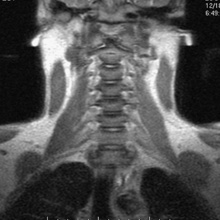
Cervical Spine Magnetic Resonance Imaging In A 30-year-old Woman With
 www.researchgate.net
cervical resonance 30year thyroid sagittal
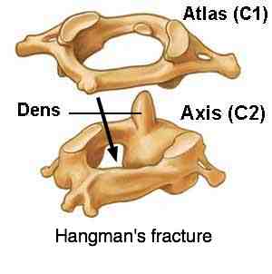
Neck Pain Anatomy Is For Those Who Want To Understand Their Problems.
 www.chiropractic-help.com
neck anatomy pain atlas axis fracture joint axial hangmans atlanto chiropractic help
Airway Imaging: Principles And Practical Guide | Anesthesia Key
 aneskey.com
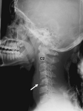
cervical spine fracture compression c5 lateral vertebra plain film demonstrates radiograph figure
Human Anatomy And Physiology Of Muscles Online On HubPages
 beth811.hubpages.com
neck muscles anatomy human bones physiology muscle lateral hubpages list whiplash cervical spine strain emedicine medscape sprain skeletal body courtesy
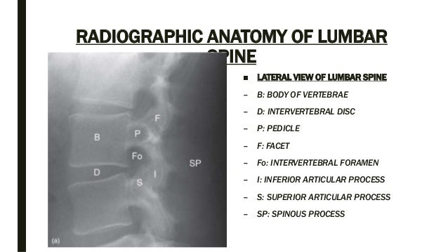
Spine Anatomy And Xray Of Spine Ppt By Dr Pratik
 www.slideshare.net
spine anatomy xray pratik
Lateral Plain Radiograph Of The Cervical Spine | The BMJ
 www.bmj.com
lateral cervical spine radiograph plain bmj
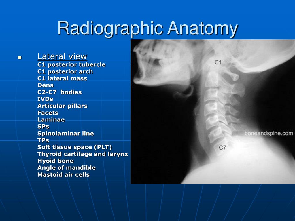
PPT - Cervical Spine PowerPoint Presentation, Free Download - ID:732240
 www.slideserve.com
c1 radiographic cervical spine mastoid arch c2 ppt powerpoint presentation posterior lateral anatomy dens
The Hyoid Bone - Human Anatomy
 www.pinterest.com
MRI Of The Spine And Bony Pelvis | Radiology Key
 radiologykey.com
spine coronal cervical mri bony pelvis figure
Cervical spine magnetic resonance imaging in a 30-year-old woman with. Neck anatomy pain atlas axis fracture joint axial hangmans atlanto chiropractic help. Lateral plain radiograph of the cervical spine