inner layer of the uterus
CARTILAGE AND BONE CELLS | Microanatomy Web Atlas | Gwen V. Childs, Ph.D.. 9 Pictures about CARTILAGE AND BONE CELLS | Microanatomy Web Atlas | Gwen V. Childs, Ph.D. : A rare case of posterior vaginal wall cyst | BMJ Case Reports, Human uterus, light micrograph - Stock Image - C020/9918 - Science and also please have a detailed explanation of the embryonic development with.
CARTILAGE AND BONE CELLS | Microanatomy Web Atlas | Gwen V. Childs, Ph.D.
 microanatomy.net
osteocytes osteoblasts cartilage microanatomy lacunae endosteum skeletal compact
A Rare Case Of Posterior Vaginal Wall Cyst | BMJ Case Reports
 casereports.bmj.com
vaginal cyst bmj casereports
Human Uterus, Light Micrograph - Stock Image - C020/9918 - Science
 www.sciencephoto.com
uterus micrograph
Fallopian Tube
 www.gfmer.ch
histology fallopian tube tubal mucosa layers internal
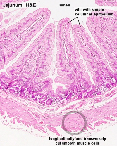
Smooth Muscle Histology - Embryology
 embryology.med.unsw.edu.au
muscle smooth histology embryology tissue layers tract circular layer gastrointestinal longitudinal outer inner colon mucosa muscularis development
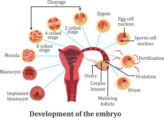
Please Have A Detailed Explanation Of The Embryonic Development With
 www.topperlearning.com
embryonic germ explanation diagrammatic topperlearning
Intrauterine Adhesions - Phoenix, AZ: Gondra Center For Reproductive
 gondracenter.com
asherman syndrome hsg adhesions uterine gynecology intrauterine uterus file hysterosalpingogram causes reproductive infertility center gondra advanced care cervical factor
Endometrium - Bloody Marvellous
 bloody-marvellous.com
endometrium uterus myometrium fallopian marvellous
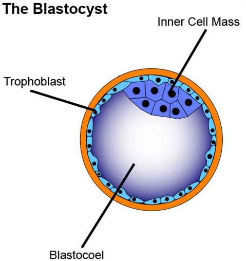
Showtime For Stem Cells
 www.independent.com
cells blastocyst stem cell embryonic mass inner blastocoel trophoblast sphere human blastocysts showtime cavity isolated
Fallopian tube. Asherman syndrome hsg adhesions uterine gynecology intrauterine uterus file hysterosalpingogram causes reproductive infertility center gondra advanced care cervical factor. Osteocytes osteoblasts cartilage microanatomy lacunae endosteum skeletal compact