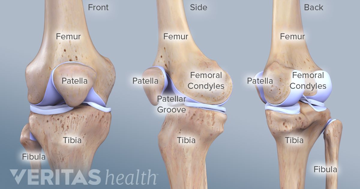
knee cap anatomy
Normal Knee X-rays | Bone and Spine. 9 Images about Normal Knee X-rays | Bone and Spine : Lab 3 27 | Chandler Physical Therapy, How Age Affects Your Risk for a Meniscus Tear and also Posterior knee dislocation - Radiology at St. Vincent's University Hospital.
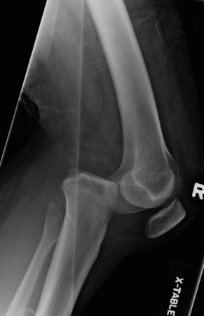
Normal Knee X-rays | Bone And Spine
 boneandspine.com
tibial limb radiographic
Transient Lateral Patellar Dislocation - Radsource
 radsource.us
patellar lateral dislocation mri radsource transient
How Age Affects Your Risk For A Meniscus Tear
 www.sports-health.com
knee bones age meniscus
Knee X-rays - Don't Forget The Bubbles
 dontforgetthebubbles.com
knee ray interpretation rays wikiradiography dontforgetthebubbles
PATELLA SUNRISE X RAY | Buyxraysonline
 buyxraysonline.com
patella buyxraysonline radiography wikiradiography
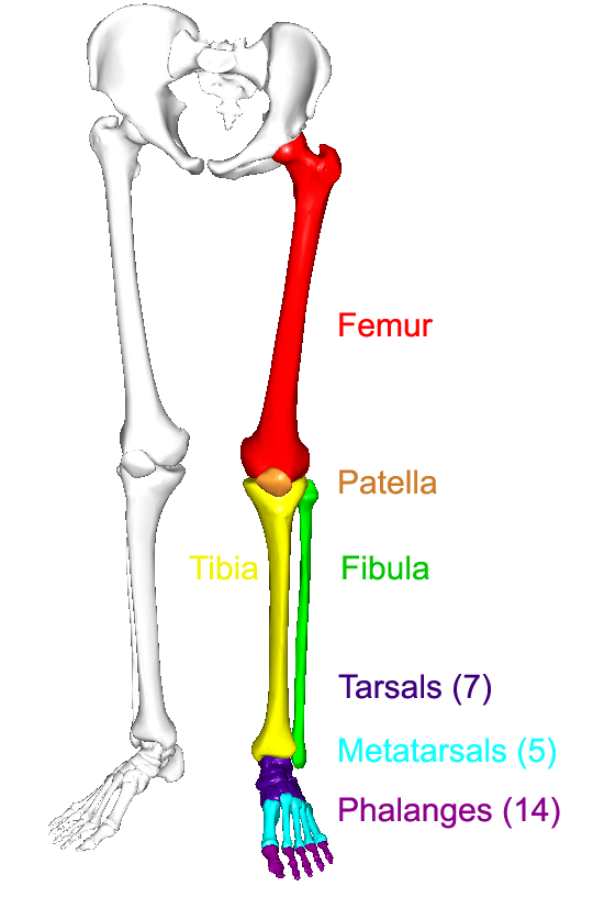
The Lower Limbs | Human Anatomy And Physiology Lab (BSB 141)
 courses.lumenlearning.com
lower limb left bones anatomy limbs human lab physiology bsb figure
Posterior Knee Dislocation - Radiology At St. Vincent's University Hospital
 www.svuhradiology.ie
dislocation knee posterior case radiology
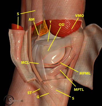
Applied Anatomy Of Knee Joint | Epomedicine
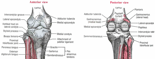 epomedicine.com
knee anatomy joint applied epomedicine tubercle medial adductor epicondyle bones muscles patella osteology condyles medical
Lab 3 27 | Chandler Physical Therapy
 chandlerphysicaltherapy.net
knee patella pain chondromalacia cap anatomy patellar labeled treatment lower tendonitis lab quadriceps muscle joint femur chiropractor tibia az chandler
Applied anatomy of knee joint. Patella sunrise x ray. Transient lateral patellar dislocation