knee joint anatomy and physiology
The tibial and femoral condyles are covered by articular cartilage and. 9 Pics about The tibial and femoral condyles are covered by articular cartilage and : Knee Joint - Part 1 - 3D Anatomy Tutorial - YouTube, CrossFit | Knee Musculature, Part 2: Posterior Muscles and also Anatomy of Shoulder | JointSurgery.in.
The Tibial And Femoral Condyles Are Covered By Articular Cartilage And
 in.pinterest.com
labelled articular sagittal tibial femur femoral femoris condyles covered quadriceps structure menisci
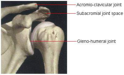
Anatomy Of Shoulder | JointSurgery.in
 www.jointsurgery.in
anatomy shoulder joint space humeral between acromio ac subacromial articulation gleno arthroscopy clavicular bony
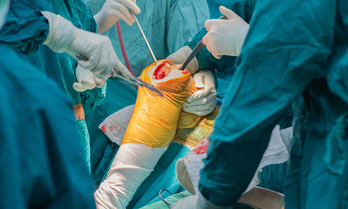
Selecting And Optimising Patients For Total Knee Arthroplasty | The
 www.mja.com.au
replacement knee total education joint care bones arthroplasty patients postoperative program plan
Radiological Anatomy Of The Lower Limb | Medical Radiography, Radiology
 www.pinterest.com
knee anatomy lateral ray joint radiology imaios radiography lower radiological medical google diagram limb bone patella radiogram rad body conventional
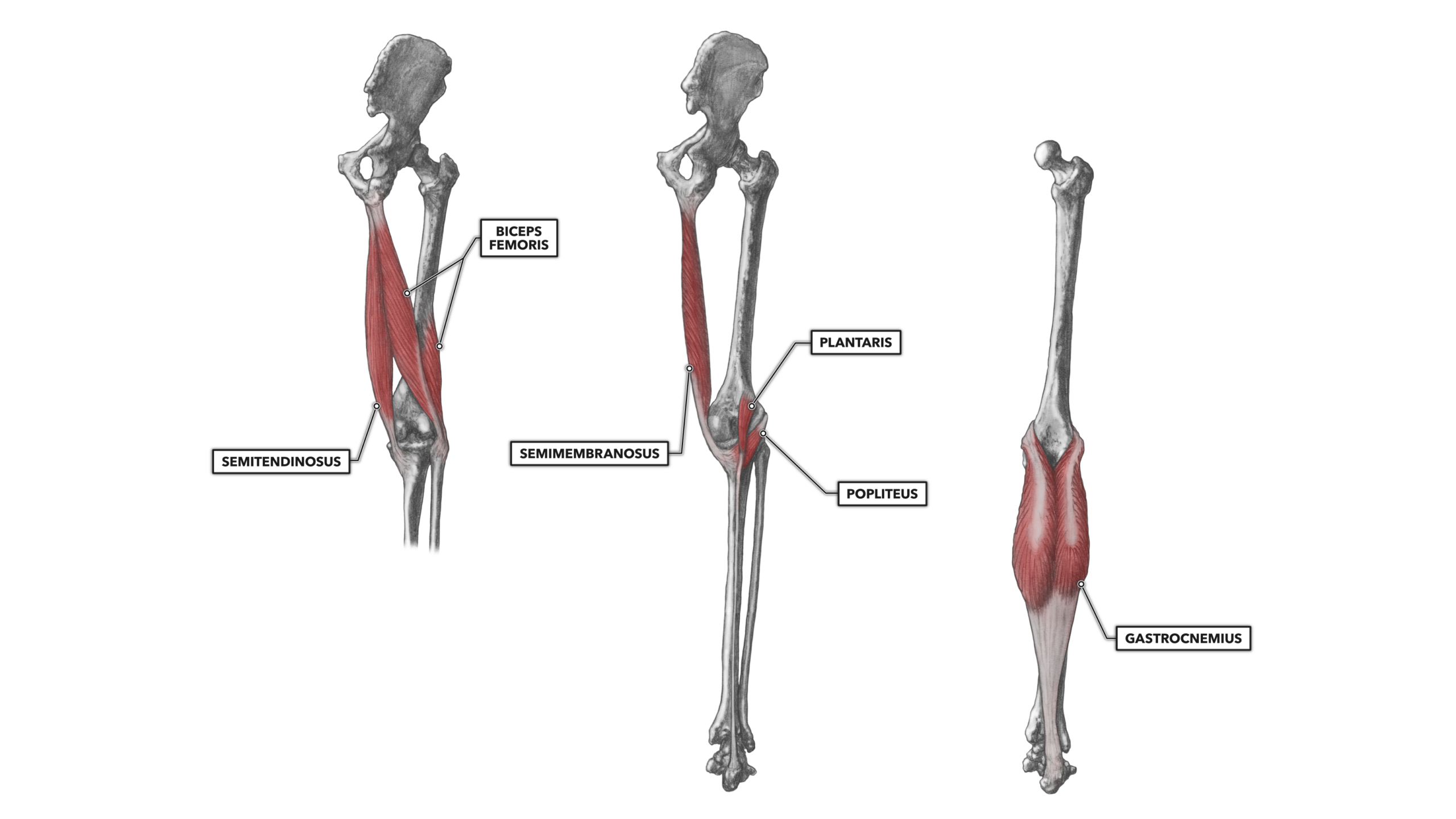
CrossFit | Knee Musculature, Part 2: Posterior Muscles
 www.crossfit.com
posterior musculature
Knee Joint - Part 1 - 3D Anatomy Tutorial - YouTube
 www.youtube.com
joint knee anatomy 3d nutra
Shoulder Anatomy
 www.fpnotebook.com
anatomy pain humerus shoulder bone arm skeleton ligament poster fpnotebook instructions printing restrictions axial
AP Of The Glenohumeral Joint | Radiology Schools, Radiology Student
 www.pinterest.com
xray radiology glenohumeral bones humerus humana scapula posterior unpacking mystery pain largely radiograph physiologie phase ombro tubercle joints limb radiografia
Finger Sprain
 www.orthoclinic.com.sg
anatomy finger hand physiology sprain
The tibial and femoral condyles are covered by articular cartilage and. Selecting and optimising patients for total knee arthroplasty. Radiological anatomy of the lower limb